Friday, December 07, 2007

Central Serous Retinopathy

Definition of Central Serous Retinopathy
Retinopathy is a condition in which deterioration of the retina is caused by damage to, or over-production of, the blood vessels in the retina.

Description of Central Serous Retinopathy
Central serous retinopathy is a slight accumulation of fluid in the macular region of the eye thatlies between the retinal pigment epithelium and the outer segments. A relative central scotoma (area of lost or depressed vision within the visual field surrounded by an area of less depressed or normal vision) results, but usually resolves spontaneously within a few months.

Serous means thin and watery-like serum.

Central serous retinopathy is characterized by an accumulation of transparent fluid at the posterior pole of the fundus (base), causing a circumscribed area of retinal detachment at the posterior pole.

When left alone, central serous retinopathy heals spontaneously within 4 to 8 weeks, with full recovery of visual acuity.

However, about one-third to one-half of all patients have recurrences after the first episode of the disease; 10 percent have three or more recurrences.

In almost half of the patients, the recurrence is within one year of the primary episode, but relapses may occur up to ten years later.

Causes and Risk Factors of Central Serous Retinopathy
Central serous retinopathy is due to a leakage, of unknown origin, through the pigment epithelium.

Symptoms of Central Serous Retinopathy
Many patients first notice a minor blurring of vision, followed by various degrees of:

* metamorphopsia (defective, distorted vision)

* micropsia (distorted visual perception in which objects appear smaller than their actual size)

* chromatopsia (visual defect in which objects appear unnaturally colored)

* central scotoma

* increasing hyperopia (farsightedness)

Visual acuity in the acute stage may range from 20/20 to 20/200 and averages 20/30. In some patients the onset of symptoms is preceded or accompanied by migraine-like headaches.

Diagnosis of Central Serous Retinopathy
The diagnosis is made by an eye examination, sometimes using a fundus contact lens.

The diagnosis is confirmed by fluorescein angiography. Typically the fluorescein enters into the blister and stains its contents, identifying one or more leakage points.

Treatment of Central Serous Retinopathy
Although no medication has thus far proved effective in treating central serous retinopathy, a beneficial effect of laser photocoagulation has been reported in several studies. Findings indicate that direct photocoagulation of the leakage point not only shortens the acute phase of the disease but also lowers the recurrence rate to about one fifth of what would be expected without active treatment.

Labels:

Tuesday, December 04, 2007

AGE-RELATED MACULAR EDEMA

Macular degeneration is a medical condition predominantly found in elderly adults in which the center of the inner lining of the eye, known as the macula area of the retina, suffers thinning, atrophy, and in some cases bleeding. This can result in loss of central vision, which entails inability to see fine details, to read, or to recognize faces. According to the American Academy of Ophthalmology, it is the leading cause of central vision loss (blindness) in the United States today for those over the age of fifty years.[1] Although some macular dystrophies that affect younger individuals are sometimes referred to as macular degeneration, the term generally refers to age-related macular degeneration (AMD or ARMD).

Age-related macular degeneration begins with characteristic yellow deposits in the macula (central area of the retina which provides detailed central vision) called drusen between the retinal pigment epithelium and the underlying choroid. Most people with these early changes (referred to as age-related maculopathy) have good vision. People with drusen can go on to develop advanced AMD. The risk is considerably higher when the drusen are large and numerous and associated with disturbance in the pigmented cell layer under the macula. Recent research suggests that large and soft drusen are related to elevated cholesterol deposits and may respond to cholesterol lowering agents or the Rheo Procedure.

Advanced AMD, which is responsible for profound vision loss, has two forms: dry and wet. Central geographic atrophy, the dry form of advanced AMD, results from atrophy to the retinal pigment epithelial layer below the retina, which causes vision loss through loss of photoreceptors (rods and cones) in the central part of the eye. While no treatment is available for this condition, vitamin supplements with high doses of antioxidants, lutein and zeaxanthin, have been demonstrated by the National Eye Institute and others to slow the progression of dry macular degeneration and in some patients, improve visual acuity.

Neovascular or exudative AMD, the wet form of advanced AMD, causes vision loss due to abnormal blood vessel growth in the choriocapillaries, through Bruch's membrane, ultimately leading to blood and protein leakage below the macula. Bleeding, leaking, and scarring from these blood vessels eventually cause irreversible damage to the photoreceptors and rapid vision loss if left untreated.

Until recently, no effective treatments were known for wet macular degeneration. However, new drugs, called anti-angiogenics or anti-VEGF (anti-Vascular Endothelial Growth Factor) agents, when injected directly into the vitreous humor of the eye using a small, painless needle, can cause regression of the abnormal blood vessels and improvement of vision. The injections frequently have to be repeated on a monthly or bi-monthly basis. Examples of these agents include Lucentis, Avastin and Macugen. Only Lucentis and Macugen are FDA approved as of April 2007. Macugen has been found to have only minimal benefits in neovascular AMD and is no longer used. Worldwide, Avastin has been used extensively despite its "off label" status. The cost of Lucentis is approximately $2000 US while the cost of Avastin is approximately $150.

[edit] Risk factors

* Aging: Approximately 10% of patients 66 to 74 years of age will have findings of macular degeneration. The prevalence increases to 30% in patients 75 to 85 years of age.[citation needed]
* Smoking: The only environmental exposure clearly associated with macular degeneration is tobacco smoking.[2] Exposure to cigarette smoke more than doubles the risk of macular degeneration.[citation needed]
* Family history: The lifetime risk of developing late-stage macular degeneration is 50% for people who have a relative with macular degeneration vs. 12% for people who do not have relatives with macular degeneration, i.e. a fourfold higher risk.[citation needed]
* Macular degeneration gene: The genes for the complement system proteins factor H (CFH) and factor B (CFB) have been determined to be strongly associated with a person's risk for developing macular degeneration. CFH is involved in inhibiting the inflammatory response mediated via C3b (and the Alternative Pathway of complement) both by acting as a cofactor for cleavage of C3b to its inactive form, C3bi, and by weakening the active complex that forms between C3b and factor B. C-reactive protein and polyanionic surface markers such as glycosaminoglycans normally enhance the ability of factor H to inhibit complement . But the mutation in CFH(Tyr402His) reduces the affinity of CFH for CRP and probably also alters the ability of factor H to recognise specific glycosaminoglycans. This change results in reduced ability of CFH to regulate complement on critical surfaces such as the specialised membrane at the back of the eye and leads to increased inflammatory response within the macula. In two 2006 studies at Yale Department of Epidemiology and Public Health and the Department of Ophthalmology and Visual Sciences, Moran Eye Center at the University of Utah School of Medicine, another gene that has implications for the disease, called HTRA1 (encoding a secreted serine protease), was identified. [3][4]
* Arg80Gly Variant of the Complement Protein C3 A genetic study published in the New England Journal of Medicine in 2007 showed that a certain, common mutation in the C3 gene which is a central protein of the Complement System is strongly associated with the occurrence of Age-related Macular Degeneration.[5] The authors consider their study to underscore the influence of the complement pathway in the pathogenesis of this disease.
* Hypertension: Also known as high blood pressure.
* Cardiovascular status - high cholesterol, obesity.
* High fat intake is associated with an increased risk of macular degeneration in both women and men. Fat provides about 42% of the food energy in the average American diet. A diet that derives closer to 20-25% of total food energy from fat is probably healthier. Reducing fat intake to this level means cutting down greatly on consumption of red meats and high-fat dairy products such as whole milk, cheese, and butter. Eating more cold-water fish[6] (at least twice weekly), rather than red meats, and eating any type of nuts may help macular degeneration patients.[7]
* Oxidative stress: It has been proposed that age related accumulation of low molecular weight, phototoxic, pro-oxidant melanin oligomers within lysosomes in the retinal pigment epithelium may be partly responsible for decreasing the digestive rate of photoreceptor outer rod segments (POS) by the RPE. A decrease in the digestive rate of POS has been shown to be associated with lipofuscin formation - a classic sign associated with macular degeneration.[8][9]

* Race Macular degeneration is more likely to be found in whites than in blacks.[10][11]
* Exposure to sunlight especially blue light. There is conflicting evidence as to whether exposure to sunlight contributes to the development of macular degeneration. A recent study in the British Journal of Ophthalmology on 446 subjects found that it does not.[12] High energy visible light (HEV) has been implicated as a cause of age-related macular degeneration.[13][14]

[edit] Signs

* Drusen
* Pigmentary alterations
* Exudative changes: hemorrhages in the eye, hard exudates, subretinal/sub-RPE/intraretinal fluid
* Atrophy: incipient and geographic
* Visual acuity drastically decreasing (two levels or more) ex: 20/20 to 20/80.

[edit] Symptoms
Image courtesy AgingEye Times

* Blurred vision: Those with nonexudative macular degeneration may by asymptomatic or notice a gradual loss of central vision, whereas those with exudative macular degeneration often notice a rapid onset of vision loss.
* Central scotomas (shadows or missing areas of vision)
* Distorted vision (i.e. metamorphopsia) - A grid of straight lines appears wavy and parts of the grid may appear blank. Patients often first notice this when looking at mini-blinds in their home.
* Trouble discerning colors; specifically dark ones from dark ones and light ones from light ones.
* Slow recovery of visual function after exposure to bright light

The Amsler Grid Test is one of the simplest and most effective methods for patients to monitor the health of the macula. The Amsler Grid is essentially a pattern of intersecting lines (identical to graph paper) with a black dot in the middle. The central black dot is used for fixation (a place for the eye to stare at). With normal vision, all lines surrounding the black dot will look straight and evenly spaced with no missing or odd looking areas when fixating on the grid's central black dot. When there is disease affecting the macula, as in macular degeneration, the lines can look bent, distorted and/or missing.

'Vision loss' or 'blindness' in macular degeneration refers to the loss of 'central vision' only. The peripheral vision is preserved. Blindness in macular degeneration does not mean 'inability to see light' and even with far advanced macular degeneration, the peripheral retina allows for useful vision.

The loss of central vision profoundly affects visual functioning. It is not possible, for example, to read without central vision. Pictures which attempt to depict the central visual loss of macular degeneration with a black spot do not really do justice to the devastating nature of the visual loss. This can be demonstrated by printing letters 6 inches high on a piece of paper and attempting to identify them while looking straight ahead and holding the paper slightly to the side. Most people find this surprisingly difficult to do.

Similar symptoms with a very different etiology and different treatment can be caused by Epiretinal membrane or macular puckeror leaking blood vessels in the eye..

[edit] Diagnosis

Fluorescein angiography allows for the identification and localization of abnormal vascular processes. Optical coherence tomography is now used by most ophthalmologists in the diagnosis and the followup evaluation of the response to treatment by using either Avastin or Lucentis which are injected into the vitreous of the eye at various intervals.

[edit] Prevention

The Age-Related Eye Disease Study showed that a combination of high-dose beta-carotene, vitamin C, vitamin E, and zinc can reduce the risk of developing advanced AMD by about 25 percent in those patients who have earlier but significant forms of the disease. This is the only proven intervention to decrease the risk of advanced AMD at this time. A follow up study, Age-Related Eye Disease Study 2 to study the potential benefits of lutein, zeaxanthine, and fish oil, is currently underway.

Anecortave acetate, (Retanne), is an anti-angiogenic drug that is given as an injection behind the eye (avoiding an injection directly into the eye) that is currently being studied as a potential way of reducing the risk of neovascular (or wet) AMD in high-risk patients.

Research started in 2005 has shown that intravitreal injection of Avastin (bevacizumab) can improve vision and slow down the macular degeneration. This therapy is currently being used in various centres around the world. Avastin is an immunoligic drug that prevents neovascularization. Hence it may also be effective in diabetic retinopathy. Avastin was initially used for the treatment of colorectal cancer.

A study by a neuroretinologist in the late 80's suggested that microcurrent stimulation of acupuncture points for the eye had positive effects in slowing and even stopping progression of the disease[citation needed]. This study was based on Ngok Cheng's research on the increased amounts of ATP levels in living tissue after being stimulated with microcurrent. ("The Effects of Electric Currents on ATP Generation, Protein Synthesis, and Membrane Transport in Rat Skin.")[15]

Recent studies suggest that statins, a family of drugs used for reducing cholesterol levels, may be effective in prevention of AMD, and in slowing its progression.[16]

Studies are underway at Harvard, with the goal of reducing lipofuscin accumulation. [9]

On September 10, 2007, in a 6-year study, researchers, led by John Paul SanGiovanni of the National Eye Institute, Maryland found that Lutein and zeaxanthin (nutrients in eggs, spinach and other green vegetables) protect against blindness (macular degeneration), affecting 1.2 million Americans, mostly after age 65. Lutein and zeaxanthin reduce the risk of AMD (journal Archives of Ophthalmology). Foods considered good sources of the nutrients also include kale, turnip greens, collard greens, romaine lettuce, broccoli, zucchini, corn, garden peas and Brussels sprouts.

Article from : http://en.wikipedia.org/wiki/Macular_degeneration

Labels: ,

Tuesday, November 28, 2006

Freedom Of Sleep

I found a very interesting post related to my earlier post. Thought my readers would enjoy reading it :).

Having A Good Sleep Helps You Get Rid Of Pressure

Too little sleep can lead to great problems or troubles. A significant risk factor for high blood
pressure is sleeplessness, as identified by the American Heart Association Journal Hypertension.

James E Gangoisch, PhD, lead author of the study and a postdoctoral fellow at the Columbia University Medical Center, says, "It has been known that one of the reason for hypertension is, sleeping disorders. This could be the reason for not getting enough sleep."

High blood pressure and short sleep (five hours or less) relationship is showed in first study. High blood pressure is significantly less in people who get sever hours or more of sleep than those who sleep five to six hours. A normal healthy person is advised to have at least 8 hours of good sleep.

Lack of sleep also can also result in obesity. The heart rate and blood pressure are normally low during sleep. Over a long period of time, the average work done by the heart increases in people deprived of sleep, which causes irreversible changes in the heart and blood vessels.

All in all a good night sleep is very essential for good health.

Sunday, November 26, 2006

Everything You Need To Know About Bird Flu

Bird flu can be one of the most dangerous epidemic if not taken care of at an early stage. Read more to know how to protect yourself and your family from bird flu.

What is avian influenza (bird flu)?

Avian influenza is an infection caused by avian (bird) influenza (flu) viruses. These flu viruses occur naturally among birds. Wild birds worldwide carry the viruses in their intestines, but usually do not get sick from them. However, avian influenza is very contagious among birds and can make some domesticated birds, including chickens, ducks, and turkeys, very sick and kill them.

Infection with avian influenza viruses in domestic poultry causes two main forms of disease that are distinguished by low and high extremes of virulence. The “low pathogenic” form may go undetected and usually causes only mild symptoms (such as ruffled feathers and a drop in egg production). However, the “highly pathogenic” form spreads more rapidly through flocks of poultry. This form may cause disease that affects multiple internal organs and has a mortality rate that can reach 90-100%, often within 48 hours.

How does avian influenza spread among birds?

Infected birds shed influenza virus in their saliva, nasal secretions, and feces. Susceptible birds become infected when they have contact with contaminated excretions or with surfaces that are contaminated with excretions or secretions. Domesticated birds may become infected with avian influenza virus through direct contact with infected waterfowl or other infected poultry or through contact with surfaces (such as dirt or cages) or materials (such as water or feed) that have been contaminated with the virus.

Do avian influenza viruses infect humans?

Bird flu viruses do not usually infect humans, but more than 100 confirmed cases of human infection with bird flu viruses have occurred since 1997.

How do people become infected with avian influenza viruses?

Most cases of avian influenza infection in humans have resulted from direct or close contact with infected poultry (e.g., domesticated chicken, ducks, and turkeys) or surfaces contaminated with secretions and excretions from infected birds. The spread of avian influenza viruses from an ill person to another person has been reported very rarely, and transmission has not been observed to continue beyond one person. During an outbreak of avian influenza among poultry, there is a possible risk to people who have direct or close contact with infected birds or with surfaces that have been contaminated with secretions and excretions from infected birds.

What are the symptoms of avian influenza in humans?

Symptoms of avian influenza in humans have ranged from typical human influenza-like symptoms (fever, cough, sore throat, and muscle aches) to eye infections, pneumonia, severe respiratory diseases (such as acute respiratory distress syndrome), and other severe and life-threatening complications. The symptoms of avian influenza may depend on which specific virus subtype and strain caused the infection.

How is avian influenza detected in humans?

A laboratory test is needed to confirm avian influenza in humans.
What are the implications of avian influenza to human health?
Two main risks for human health from avian influenza are 1) the risk of direct infection when the virus passes from the infected bird to humans, sometimes resulting in severe disease; and 2) the risk that the virus – if given enough opportunities – will change into a form that is highly infectious for humans and spreads easily from person to person.

How is avian influenza in humans treated?

Studies done in laboratories suggest that the prescription medicines approved for human influenza viruses should work in treating avian influenza infection in humans. However, influenza viruses can become resistant to these drugs, so these medications may not always work. Additional studies are needed to determine the effectiveness of these medicines.

Friday, November 17, 2006

PARKINSON’S DISEASE – very common among the older age group! What is it?

PARKINSON’S DISEASE is a progressive degenerative disorder, mostly affecting the older people.

Why is PARKINSON’S DISEASE called so?
James Parkinson first described it in 1817.

What are the causes for PARKINSON’S DISEASE, a very common ailment?
Well most of the times it is idiopathic (i.e. the reason is unknown). But other causes may be trauma, toxic agents, drugs (dopamine antagonists)

What are the clinical features of PARKINSON’S DISEASE ? How does it present itself?

· Diminished facial _expression
· Stooped posture
· Slowness of voluntary movement
· Festinating gait (progressively shortened, accelerated steps)
· Rigidity
· “Pill rolling” movements of the fingers
· Tremors
· Dementia may also occur

If untreated the symptoms progress over several years to end stage disease in which the patient is so rigid that he is unable to move, unable to breathe properly; succumbs mostly to chest infections/ embolism.

Wednesday, November 08, 2006

A Perfect Solution For Hair Loss Problem As a Result Of Wearing Helmet

I've always hated wearing helmet. No, not that I don't love my head or want to keep it safe. I understand the importance of wearing helmet. But it has a purpose, you too would hate to wear a helmet in a slow moving traffic, (I'm talking about traffic which moves at around 10km/hr !! ).

Recently Karnataka Government came up with a new rule stating everyone ( riders only thankfully, lucky pillionz ) must and should wear a helmet else they would be fined. Now you might call this a very crude way of making money government has come up with, getting to the purpose of this article, which is not discussing whether the decision made by government is right or wrong but is to discuss some other serious problems involved with wearing a helmet.

The most common problem everyone will face as a result of this new rule is Hair Fall. Imagine wearing those heavy helmets, travelling on a average for about an hr or more a day, and spending even more time waiting for those traffic signals to go green. Spending hours together in those "always exepected" traffic jams. None of this is going to do any good for your hair.

All the dust, wear and tear just helps your hair grow much weaker which eventually will end up with experiencing hair fall.

But by following a few steps you definitely can protect your hair from all the wear and tear and also from getting spoilt because of the Helmets you wear.

Wet a piece of cloth and wear it as a "Bandana" before you wear your helmet. This protects your hair in two ways.

One, it keeps the moisture in your hair, which is the most essential part in mainiting your hairs bounciniess and retaining life in them.
Second, it reduces the friction between your hair and the soft sponge inside your helmet. the friction between your hair and the sponge inside your helmet does not do any good for your hair.

It also becomes very important that you buy a helmet which fits you nice and tight. This also helps in reducing the friction.

Don't forget to regularly oil your hair. There is nothing like a good oil massage.

Wash and clean your hair regularly. Rinse it thoroughly. The more clean you keep them, the more healthier your hair will be.

Sunday, November 05, 2006

How Long Does It Take For Hoodia Gordonii To Work ?

Hoodia Gordonii is a succelent generally found in the Kalahari desert. It is believed to have hunger suppressing powers.

Nutrition and Diet is one of the biggest industry today where thousands of people are looking for new ways to loose weight. What they actually look for is an "easy" way to loose weight and hence there is great demand for diet pills and diet syrups.

Hoodia Gordonii is a diet pill, which has been discussed regularly and is seemingly popular amongst people planning on a diet.

Generally Hoodia Gordonii takes just about half an hour to start showing it's supressing effect and this makes it extremely popular.

Individuals who plan to take diet pills, should consult their doctors before they try testing any of the diet pills available in the market. There are a lot of chances that more than 60% of those pills, don't live up to what they claim.

Source : Hoodia Gordonii

Tuesday, October 24, 2006


Tuberculosis (commonly shortened to TB) is an infection caused by the bacterium Mycobacterium tuberculosis, which most commonly affects the lungs (pulmonary TB) but can also affect the central nervous system (meningitis), lymphatic system, circulatory system (Miliary tuberculosis), genitourinary system, bones and joints.

Tuberculosis is one of the most deadly and common major infectious diseases today, infecting two billion people, or approximately one-third ( WHO TB.) of the world's population. Nine million new cases of disease, resulting in two million deaths, occur annually, mostly in developing countries. However, developed countries are not spared the burden of tuberculosis. There is a rising proportion of people who are immunocompromised, either due to immunosupressive drugs or HIV/AIDS. These people are at particular risk of tuberculosis infection and active tuberculosis disease.

Most of those infected (90%) have asymptomatic latent TB infection (LTBI). There is a 10% lifetime chance that LTBI will progress to TB disease which, if left untreated, will kill more than 50% of its victims. TB is one of the top three infectious killing diseases in the world: HIV/AIDS kills 3 million people each year, TB kills 2 million, and malaria kills 1 million.

The neglect of TB control programs, HIV/AIDS, and immigration has caused a resurgence of tuberculosis. Multiple drug resistant strains of TB (MDR-TB) and Extreme Drug-Resistance in Tuberculosis (XDR-TB) are emerging. The World Health Organization declared TB a global health emergency in 1993, and the Stop TB Partnership proposed a Global Plan to Stop Tuberculosis which aims to save an additional 14 million lives between 2006 and 2015.

Other names for the disease

TB (short for tuberculosis and also for Tubercle Bacillus)

Consumption (TB seemed to consume people from within with its symptoms of bloody cough, fever, pallor, and long relentless wasting)

Wasting disease

White plague (TB sufferers appear markedly pale)

Phthisis (Greek for consumption) and phthisis pulmonalis

Scrofula (swollen neck glands)

King's evil (so called because it was believed that a king's touch would heal scrofula)

Pott's disease of the spine

Miliary TB (x-ray lesions look like millet seeds)

Tabes mesenterica (TB of the abdomen)

Lupus vulgaris (the common wolf - TB of the skin)

Prosector's wart, also a kind of TB of the skin, transmitted by contact with contaminated cadavers to anatomists, pathologists, veterinarians, surgeons, butchers, etc.

Koch's Disease named after Robert Koch who discovered the tuberculosis bacilli.

The bacterium

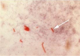
Acid-fast bacilli (AFB) (shown in red) are tubercle bacilli Mycobacterium tuberculosis.

The cause of tuberculosis, Mycobacterium tuberculosis (MTB), is a slow-growing aerobic bacterium that divides every 16 to 20 hours. This is extremely slow compared to other bacteria (although not the slowest), which tend to have division times measured in minutes (among the fastest growing bacteria is a strain of E. coli that can divide roughly every 20 minutes; by contrast, Mycobacterium leprae divides every 20 days). MTB is not classified as either Gram-positive or Gram-negative because it does not have the chemical characteristics of either, although it contains peptidoglycan in the cell wall, which means it is related to the Gram-positive organisms. If a Gram stain is performed, it stains very weakly Gram-positive or not at all (ghost cells). It is a small rod-like bacillus which can withstand weak disinfectants and can survive in a dry state for weeks but, spontaneously, can only grow within a host organism (in vitro culture of M. tuberculosis took a long time to be achieved, but is nowadays a routine laboratory procedure).

MTB is identified microscopically by its staining characteristics: it retains certain stains after being treated with acidic solution, and is thus classified as an "acid-fast bacillus" or "AFB". In the most common staining technique, the Ziehl-Neelsen stain, AFB are stained a bright red which stands out clearly against a blue background. Acid-fast bacilli can also be visualized by fluorescent microscopy, and by an auramine-rhodamine stain.

The M. tuberculosis complex includes 3 other mycobacteria which can cause tuberculosis: M. bovis, M. africanum, and M. microti. The first two are very rare causes of disease and the last one does not cause human disease.

Nontuberculous mycobacteria (NTM) are other mycobacteria (besides M. leprae which causes leprosy) which may cause pulmonary disease resembling TB, lymphadenitis, skin disease, or disseminated disease. These include Mycobacterium avium, M. kansasii, and others.

The disease Transmission

TB is spread by aerosol droplets expelled by people with active TB disease of the lungs when they cough, sneeze, speak, or spit. Each droplet is 5 µm in diameter and contains 1 to 3 bacilli. Close contacts (people with prolonged, frequent, or intense contact) are at highest risk of becoming infected (typically a 22% infection rate). A person with untreated, active tuberculosis can infect an estimated 20 other people per year. Others at risk include foreign-born from areas where TB is common, immunocompromised patients (eg. HIV/AIDS), residents and employees of high-risk congregate settings, health care workers who serve high-risk clients, medically underserved, low-income populations, high-risk racial or ethnic minority populations, children exposed to adults in high-risk categories, and people who inject illicit drugs.

Transmission can only occur from people with active TB disease (not latent TB infection).

The probability of transmission depends upon infectiousness of the person with TB (quantity expelled), environment of exposure, duration of exposure, and virulence of the organism.

The chain of transmission can be stopped by isolating patients with active disease and starting effective anti-tuberculous therapy.

Pathogenesis

While only 10% of TB infection progresses to TB disease, if untreated the death rate is 51%.

TB infection begins when MTB bacilli reach the pulmonary alveoli, infecting alveolar macrophages, where the mycobacteria replicate exponentially. The primary site of infection in the lungs is called the Ghon focus. Bacteria are picked up by dendritic cells, which can transport the bacilli to local (mediastinal) lymph nodes, and then through the bloodstream to the more distant tissues and organs where TB disease could potentially develop: lung apices, peripheral lymph nodes, kidneys, brain, and bone.

Tuberculosis is classed as one of the granulomatous inflammatory conditions. Macrophages, T lymphocytes, B lymphocytes and fibroblasts are among the cells that aggregate to form a granuloma, with lymphocytes surrounding infected macrophages. The granuloma functions not only to prevent dissemination of the mycobacteria, but also provides a local environment for communication of cells of the immune system. Within the granuloma, T lymphocytes (CD4+) secrete a cytokine such as interferon gamma, which activates macrophages to destroy the bacteria with which they are infected, making them better able to fight infection. T lymphocytes (CD8+) can also directly kill infected cells.

Importantly, bacteria are not eliminated with the granuloma, but can become dormant, resulting in a latent infection. Latent infection can be diagnosed only by tuberculin skin test, which yields a delayed hypertype sensitivity response to purified protein derivatives of M. tuberculosis in an infected person.

Another feature of the granulomas of human tuberculosis is the development of cell death, also called necrosis, in the center of tubercles. To the naked eye this has the texture of soft white cheese and was termed caseous necrosis.

If TB bacteria gain entry to the blood stream from an area of tissue damage they spread through the body and set up myriad foci of infection, all appearing as tiny white tubercles in the tissues. This is called miliary tuberculosis and has a high case fatality.

In many patients the infection waxes and wanes. Tissue destruction and necrosis are balanced by healing and fibrosis. Affected tissue is replaced by scarring and cavities filled with cheese-like white necrotic material. During active disease, some of these cavities are in continuity with the air passages bronchi. This material may therefore be coughed up. It contains living bacteria and can pass on infection.

Treatment with appropriate antibiotics kills bacteria and allows healing to take place. Affected areas are eventually replaced by scar tissue.

Progression

In those people in whom TB bacilli overcome the immune system defenses and begin to multiply, there is progression from TB infection to TB disease. This may occur soon after infection (primary TB disease – 1 to 5%) or many years after infection (post primary TB, secondary TB, reactivation TB disease of dormant bacilli – 5 to 9%). The risk of reactivation increases with immune compromise, such as that caused by infection with HIV. In patients co-infected with M. tuberculosis and HIV, the risk of reactivation increases to 10% per year, while in immune competent individuals, the risk is between 5 and 10% in a lifetime.

About five percent of infected persons will develop TB disease in the first two years, and another five percent will develop disease later in life. In all, about 10% of infected persons with normal immune systems will develop TB disease in their lifetime.

Some medical conditions increase the risk of progression to TB disease. In HIV infected persons with TB infection, the risk increases to 10% each year instead of 10% over a lifetime. Other such conditions include drug injection (mainly because of the life style of IV Drug users), substance abuse, recent TB infection (within two years) or history of inadequately treated TB, chest X-ray suggestive of previous TB (fibrotic lesions and nodules), diabetes mellitus, silicosis, prolonged corticosteroid therapy and other immunosuppressive therapy, head and neck cancers, hematologic and reticuloendothelial diseases (leukemia and Hodgkin's disease), end-stage renal disease, intestinal bypass or gastrectomy, chronic malabsorption syndromes, or low body weight (10% or more below the ideal).

Some drugs, including rheumatoid arthritis drugs that work by blocking tumor necrosis factor-alpha (an inflammation-causing cytokine), raise the risk of causing a latent infection to become active due to the importance of this cytokine in the immune defense against TB.

TB disease most commonly affects the lungs (75% or more), where it is called pulmonary TB. Symptoms include a productive, prolonged cough of more than three weeks duration, chest pain, and hemoptysis. Systemic symptoms include fever, chills, night sweats, appetite loss, weight loss, and easy fatigability. The term consumption arose because sufferers appeared as if they were "consumed" from within by the disease. People from Asian and African descent may have more often lymph node TB than Caucasians.

Extrapulmonary sites include the pleura, central nervous system (meningitis), lymphatic system (scrofula of the neck), genitourinary system, and bones and joints (Pott's disease of the spine). An especially serious form is "disseminated", or "miliary" TB, so named because the lung lesions so-formed resemble millet seeds on x-ray. These are more common in immunosuppressed persons and in young children. Pulmonary TB may co-exist with extrapulmonary TB.

Drug resistance

Drug-resistant TB is transmitted in the same way as regular TB. Primary resistance develops in persons initially infected with resistant organisms. Secondary resistance (acquired resistance) may develop during TB therapy due to inadequate treatment regimen, not taking the prescribed regimen appropriately or using low quality medication.

Diagnosis

A complete medical evaluation for TB includes a medical history, a physical examination, a tuberculin skin test, a serological test, a chest X-ray, and microbiologic smears and cultures. The measurement of a positive skin test depends upon the person's risk factors for progression of TB infection to TB disease. Bacteriophage-based assays are among a few new testing procedures that offer the hope of cheap, fast and accurate TB testing for the impoverished countries that need it most.

See: tuberculosis diagnosis, tuberculosis radiology

Treatment

Persons with TB infection (class 2 or class 4 TB), but who do not have TB disease (class 3 or class 5 TB), cannot spread the infection to other people. TB infection in a person who does not have TB disease is not considered a case of TB and is often referred to as latent TB infection (LTBI). This distinction is important because treatment options will be different for a person who has LTBI instead of active TB disease.

See: tuberculosis treatment

Prevention of Tuberculosis

Prevention and control efforts include three priority strategies:

identifying and treating all persons who have TB disease

finding and evaluating persons who have been in contact with TB patients to determine whether they have TB infection or disease, and treating them appropriately, and

testing high-risk groups for TB infection to identify candidates for treatment of latent infection and to ensure the completion of treatment.

In tropical areas where the incidence of atypical mycobacteria is high, exposure to nontuberculous mycobacteria gives some protection against TB.

BCG vaccine

Many countries use BCG vaccine as part of their TB control programs, especially for infants. The protective efficacy of BCG for preventing serious forms of TB (e.g. meningitis) in children is high (greater than 80%). However, the protective efficacy for preventing pulmonary TB in adolescents and adults is variable, from 0 to 80%. In the United Kingdom, children aged 10-14 were typically immunized during school until 2005. (Routine BCG vaccination was stopped as it was no longer cost-effective. The incidence of TB in people born in the UK, and with parents and grandparents who were born in the UK, was at an all time low, and falling. Others continue to be offered BCG vaccination.)

The effectiveness of BCG is much lower in areas where mycobacteria are less prevalent. In the USA, BCG vaccine is not routinely recommended except for selected persons who meet specific criteria:

Infants or children with negative skin-test result who are continually exposed to untreated or ineffectively treated patients or will be continually exposed to multi-drug resistant TB.

Healthcare workers considered on individual basis in settings in which high percentage of MDR-TB patients has been found, transmission of MDR-TB is likely, and TB control precautions have been implemented and not successful.

Tuberculosis vaccine

The first recombinant tuberculosis vaccine entered clinical trials in the United States in 2004 sponsored by the National Institute of Allergy and Infectious Diseases (NIAID). [1]

A 2005 study showed that a DNA TB vaccine given with conventional chemotherapy can accelerate the disappearance of bacteria as well as protecting against re-infection in mice; it may take four to five years to be available in humans. PMID 15690060.

Because of the limitations of current vaccines, researchers and policymakers are promoting new economic models of vaccine development including prizes, tax incentives and advance market commitments

Monday, September 18, 2006

TUBERCULOSIS and modes of Transmission

TUBERCULOSIS

Tissue response in tuberculosis represents classical example of chronic granulomatous inflammation in humans.the causative agent is tubercle bacillus or Koch s bacillus called Mycobacterium tuberculousis causes tuberculosis in the lung and other tissues of the human body. The organism is a strict aerobe and thrives best in tissues with oxygen tension like in the apex of the lung.

Mode Of Transmission

  1. Inhalation of organisms present in fresh cough droplets or in dried sputum form an open case of pulmonary tuberculosis.
  2. Ingestion of the organisms leads to development of tonsillar or intestinal tuberculosis. This mode of infection of human tubercle bacilli is from self-swallowing of infected sputum of an open case of pulmonary tuberculosis, or ingestion of bovine tubercle bacilli from milk of diseased cows.
  3. Inoculation of the organisms into the skin may rarely occur from infected postmorten tissue.
  4. transplacental route results in development of congenital tuberculosis in foetus from infected mother and is a rare mode of transmission.

Friday, August 18, 2006

STROKE: Remember The 1st Three Letters...
A Stroke can be really dangerous if you don't realize you've had one at the right time. Make sure you know these points so that you are safe, no matter what
STROKE IDENTIFICATION:


During a BBQ, a friend stumbled and took a little fall - she assured everyone that she was fine (they offered to call paramedics) and just tripped over a brick because of her new shoes. They got her cleaned up and got her a new plate of food - while she appeared a bit shaken up, Ingrid went about enjoying herself the rest of the evening. Ingrid's husband called later telling everyone that his wife had been taken to the hospital - (at
6:00pm, Ingrid passed away.) She had suffered a stroke at the BBQ. Had they known how to identify the signs of a stroke, perhaps Ingrid would be with us today. Some don't die. They end up in a helpless, hopeless condition instead.


It only takes a minute to read this...

A neurologist says that if he can get to a stroke victim within 3 hours he can totally reverse the effects of a stroke...totally. He said the trick was getting a stroke recognized, diagnosed, and then getting the patient medically cared for within 3 hours, which is tough.


RECOGNIZING A STROKE

Thank God for the sense to remember the "3" steps, STR . Read and Learn!

Sometimes symptoms of a stroke are difficult to identify. Unfortunately, the lack of awareness spells disaster. The stroke victim may suffer severe brain damage when people nearby fail to recognize the symptoms of a stroke.
Now doctors say a bystander can recognize a stroke by asking three simple questions:

S
*Ask the individual to SMILE.
T
*Ask the person to TALK . to SPEAK A SIMPLE SENTENCE
(Coherently) (i.e. . . It is sunny out today)
R
*Ask him or her to RAISE BOTH ARMS .
{NOTE: Another 'sign' of a stroke is this: Ask the person to 'stick' out their tongue... if the tongue is 'crooked', if it goes to one side or the other that is also an indication of a stroke}

If he or she has trouble with ANY ONE of these tasks, call for assistance immediately and describe the symptoms to the dispatcher.

Tuesday, August 15, 2006

BRONCHOGENIC CARCINOMA or lung cancer

BRONCHOGENIC CARCINOMA :

A number of benign and malignant tumours occur in the lungs but the primary lung cancer, commonly termed bronchogenic carcinoma, is the most common. The lung is also the commonest site for metastasis from carcinomas and sarcomas. A histologic classification of various benign and malignant tumours of lungs as recommended by the World Health Organisation

It is the most common primary malignant tumour in men inindustrialised nations and accounts for nearly one-third of all cancer deaths in both sexes. Currently, the incidence in females in the United States has already exceeded breast cancer as a cause of death in women. Cancer of the lung is a disease of middle and late life with peak incidence in 5th to 7th decades, after which there is gradual fall in its incidence.

ETIOPATHOGENESIS

1. Smoking
2. Atmospheric pollution
3. Occupational causes
4. Dietary factors
5. Genetic factors
6. Chronic Scarring

Thursday, August 03, 2006

Hoodia is considered a Magic Pill in the Weight Loss Industry

Hoodia Gordonii is a cactus found in the Kalahari Desert. The use of Hoodia has been long known by the indigenous populations of Southern Africa, who infrequently use these plants for treating indigestion and small infections.
But scientifically Hoodia Gordonii has an ingredient called P57 which is used in treating obesity. The molecular P57 is believed to be responsible for curbing ones appetite.
But consumers should be careful in picking the authentic hoodia from the market as there seems to be a lot of companies which are illegal.
So far, there is no negative report against hoodia. Hoodia is considered to be safe but its always a good practice to consult your doctor before picking up hoodia.
For those who are concerned about reducing weight, my serious advice is to learn tactics to speed up your metabolism rate. Its a healthier and cleaner way to loose weight.

Wednesday, July 26, 2006

What is Anaemia ??

Anaemia… wat is it?

Anaemia is defined as haemoglobin concentration in blood below the lower limit of the normal range for the age and sex of the individual. In adults, the lower extreme of the normal haemoglobin is taken as 13 g% for males and 11.5% for females. New born infants have higher haemoglobin levels and therefore, 15% is taken as the lowr limit at birth, whereas at 3 months , the lower limit is taken as 9.5%. Although haemoglobin value is employed as the only parameter for determining whether or not anaemia is present, the red cell counts, haematocrit(PCV) and absolute values (MCV, MCH and MCHC) provide alternate means of assessing anaemia.

What are the symptoms of anaemia?
· Tiredness
· Easy fatiguability
· Generalized muscular weakness
· Lethargy
· Headache
· In older patients- there maybe symptoms of cardiac failure, angina pectoris, intermittent claudication, confusion and visual disturbances.

What actually happens in an anaemic person?

Subnormal level of haemoglobin causes lowered oxygen- carrying capacity of the blood. This in turn, initiates compensatory physiologic adaptations such as:
· Increased release of oxygen from haemoglobin;
· Increased blood flow to the tissues;
· Maintainance of the blood volume; and
· Redistribution of blood flow to maintain the cerebral blood supply.
Eventually, however, tissue hypoxia (lack of oxygen) develops causing impaired functions of the affected tissues. The degree of functional impairment of individual tissues is variable depending upon their oxygen requirements. Tissues with high oxygen requirement such as the heart, CNS and the skeletal muscle during exercise, bear the brunt of clinical effects of anaemia

Sunday, July 02, 2006

What is Mesothelioma

Mesothelioma is an uncommon tumour arising from mesothelial lining of serous cavities, most often in the pleural (covering of lungs) cavity, and rarely in peritoneal (coverings of abdomen) cavity and pericardial (covering of the heart) sac. They are of two types --- benign (solitary) and malignant (diffuse). The biological behavior of the mesothelioma’s can be usually predicted by their gross appearance; thoses forming solitary, discrete masses are generally benign, whereas those, which grow diffusely, are usually malignant.
BENIGN (SOLITARY) MESOTHELIOMA
Benign or solitary mesothelioma is also called pleural fibroma. Asbestos exposure plays no role on the etiology of benign mesothelioma. Grossly it consists of of a solitary, circumscribed, small, firm mass, generally less than 3cm in diameter. Cut surface shows whorls of dense fibrous tissue. Microscopically, the tumour is predominantly composed of whorls of collagen fibres and reticulin with interspersed fibroblasts. Rarely, mesothelial-lined clefts are seen in the tumour. Benign mesothelioma causes no symptoms and is detected as an incidental radiologic finding. Sometimes the tumour is associated with systemic syndrome of osteoarthropathy or hypoglycemia. Removal of the tumour is generally curative.

MALIGNANT (DIFFUSE) MESOTHELIOMA
Malignant or diffuse mesothelioma is rare. It is a highly malignant tumour associated with high mortality. The tumour is significant in view of its recognized association with occupational exposure to asbestos for a number of years, usually 20-40 years. About 90% of malignant mesothelioma’s are asbestos –related. Mechanism of carcinogenecity by asbestos is not quite clear but it appears that prolonged exposure of amphibole type of asbestos is capable of inducing oncogenic mutation in the mesothelium. However prolonged exposure is considered more significant rather than heavy exposure as documented by its occurrence in the family of asbestos workers.
Although combination of cigarette smoking and asbestos exposure greatly increases risk to develop bronchgenic carcinoma, there is no such extra increased risk of developing mesothelioma in asbestos workers who smoke. Recently, SV40 (simian vacuolating virus) has also been implicated in the etiology of mesothelioma.

Sunday, May 07, 2006

Congenital Heart Disease (CHD)

Very often we come across a question, What is Congenital Heart Disease ?, What are the reasons for Congeital Heart Disease ? and a few more. I'm trying to answer a few of these, if any one would want to know more, please feel free to post your questions in comments section. I'll try to answer or get the right answer for from the right sources.


What is Congenital Heart Disease ?
Congenital heart disease is the abnormality of the heart present from birth. It is the most common and important form of heart disease in the early years of life and is present in about 0.5% of newborn children.

Congenital Heart Disease is normally found in ?
This form of heart disease is normally noticed in the early years of life and is present in about 0.5% of newborn children. The incidence is higher in premature infants.

What are the causes of Congenital Heart Disease ?
The cause of congenital heart disease is unknown in most of the cases. It is attributed to multifactorial inheritance involving genetic and environmental influences.

Any other factors, which may result in Congenital Heart Disease ?
Other factors like rubella virus infection to the mother during pregnancy, drugs taken by the mother anf heavy alcohol drinking by the momther have all been implicated in causing utero injury resulting in congenital malformations of the heart.

Are there any ways of classifying Congeital Heart Disease ?
There are many ways of Classifying CHD:
1> Malformations of the Heart,
2> Shunts ( Cyanotic Congenital Heart Disease),
3>Obstructions ( obstructive congenital heart disease).